|
Welcome
Welcome to the University of Alabama at Birmingham FBS Portal. This site is designed to automate the use of our Core Facilities and to provide the best possible customer service. Quick Info
For more info, please contact the PSI Support Team. | Our Core Facilities To learn more about a particular facility or to request access, please click on a facility name below. Comprehensive Cancer CenterUniversity of Alabama at Birmingham |
Main Contact Info
| Other Contacts | |||
|---|---|---|---|
| Melissa Chimento | Associate Director | (205) 934-1926 | mchimento@uab.edu |
| Shawn R. Williams | Fluorescence Microscopy | (205) 934-7403 | tzaron@uab.edu |
| Robert Grabski | Fluorescence Microscopy | (205) 934-7309 | rgrabski@uab.edu |
| Alexa L Mattheyses | Director | mattheyses@uab.edu | |
This facility has not published any Products. Please check back.
The following Products and Services are available within our facility:
Instruments
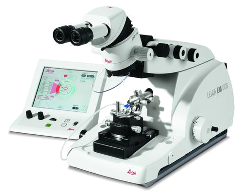 |
Cryo UltramicrotomeThe Ultramicrotome Leica EM UC6 provides easy preparation of semi- and ultrathin sections as well as perfect, smooth surfaces of biological and industrial samples for TEM, SEM, AFM and LM examination. |
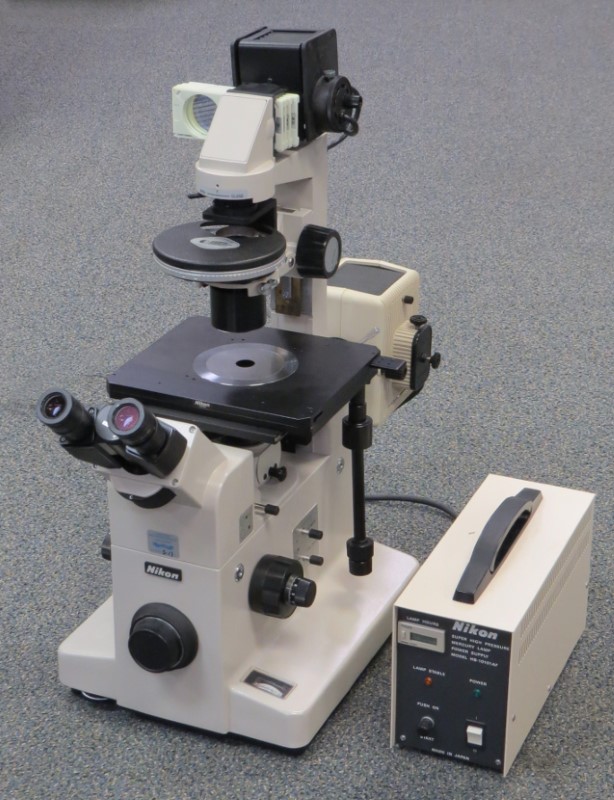 |
DiaphotNikon Diaphot 300 Inverted Fluorescence Phase contrast microscope. |
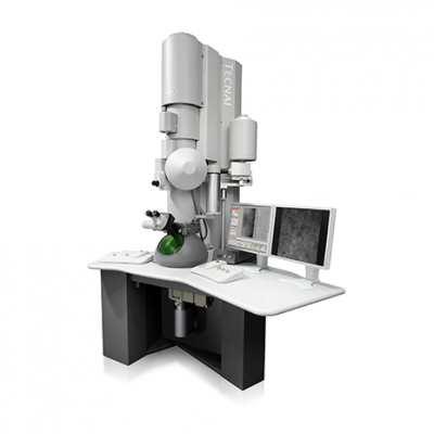 |
FEI Tecnai T12 - TEMThe Tecnai Spirit T12 Transmission Electron Microscope (Thermo-Fisher, formerly FEI) was installed the basement of the Shelby Biomedical Building in 2006 and has been operational since April of 2007. It has an operating voltage range of 20 to 120 kV and a magnification range of 18.5x-650kx. |
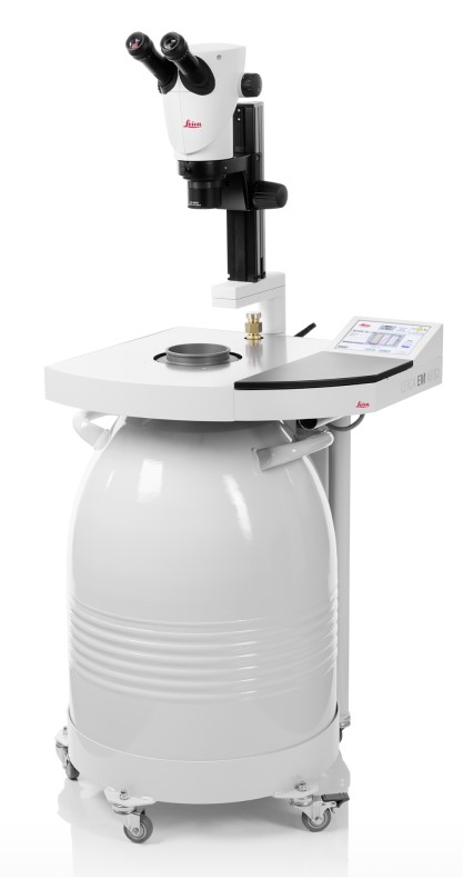 |
Freeze Substitution System (AFS)Freeze-substitution is a process of dehydration, performed at temperatures low enough to avoid the formation of ice crystals and to circumvent the damaging effects observed after ambient-temperature dehydration. During freeze substitution the "frozen" water is dissolved by an organic solvent, which usually also contains chemical fixatives [1]. |
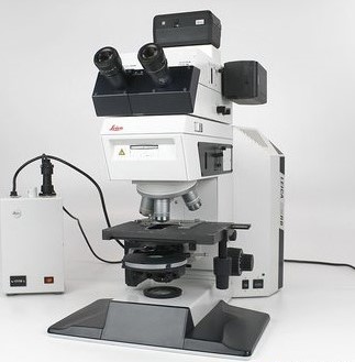 |
Leica DMRBThe Leica Leitz DM RB microscope provides enhanced object identification by using fluorescence illumination. It also allows bright and darkfield illumination techniques, phase contrasts, and condenser height adjustment. |
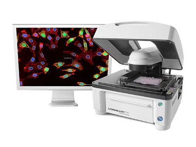 |
Lionheart FXThe Lionheart is an Automated Microscope System that can Image in four modes, Bright Field (B&W) Color Brightfield, Phase and Fluorescence. Capable of stitching Color Brightfield and Fluorescent images along with Augmented Advanced Analysis software. |
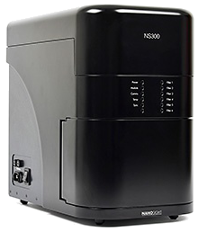 |
Nanosight NS 300The Nanosight NS 300 is used to image small particles below the micron threshold. For looking at Exosomes and other small particles that are too small for a confocal or flow cytometry. |
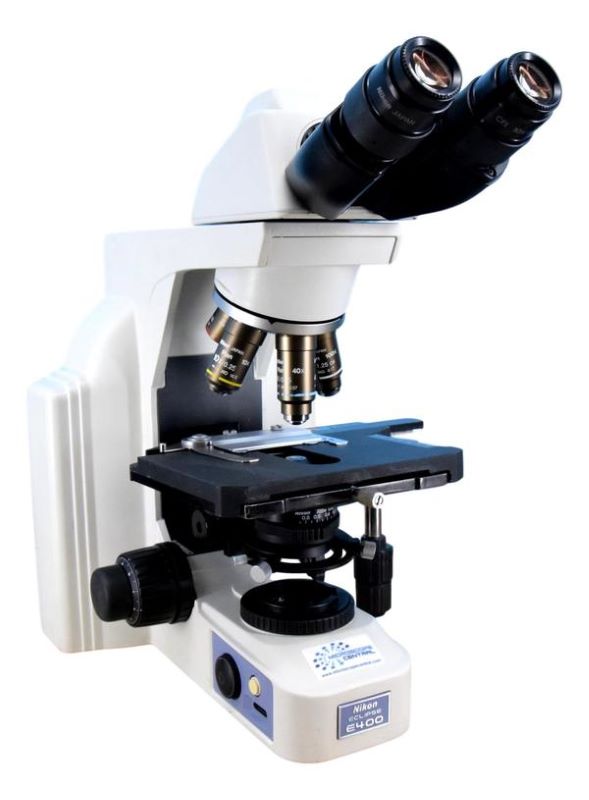 |
Nikon EclipseThe Nikon Eclipse microscope is ideal for clinical applications. It is a popular microscope for routine biological applications, basic Imaging, Timelapse and Advanced Densitometry. |
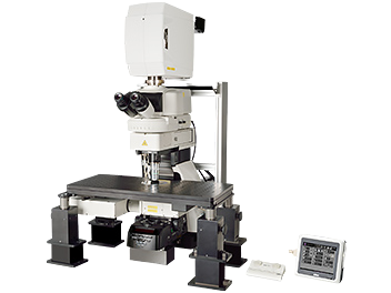 |
Nikon Multiphoton ConfocalThe Nikon Multiphoton A1R Laser Confocal Microscope is a specialized instrument used for imaging live animals and tissue which have been treated with fluorescent probes. |
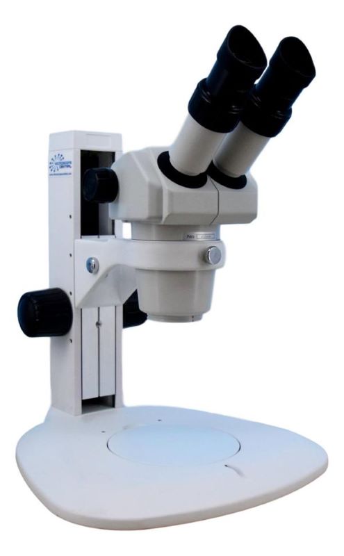 |
Nikon StereoThe Nikon Binocular Stereo Zoom Microscope delivers outstanding optical performance using Porro prisms that provide a lightweight, compact design. |
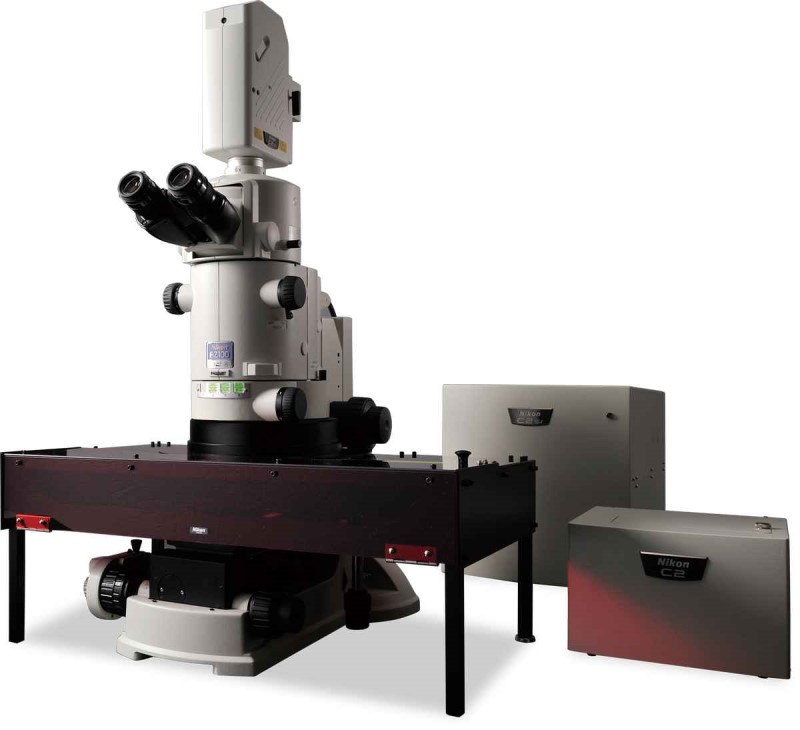 |
Nikon Storm C2siNikon STORM Super Resolution Confocal Imaging System. It has STORM, TIRF, and a C2si Scan Head with DUVB Scanner Spectral Detection. |
 |
Nis-Analysis PCAdvanced Nis Elements Analysis PC Workstation and Imaging Analysis Computer can be used for saving pictures from raw Nis Elements .nd2 data files. It also has FIJI, Mattlab and several other imaging packages for analysis and manipulating image data. |
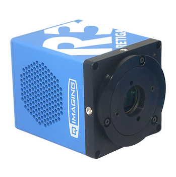 |
Q-Imaging CameraQ-Imaging Digital Camera is for use with all microscopes in Shelby 136A. |
Remember to acknowledge the HRIF core in your publications:
General: "Research reported in this publication was supported by the UAB High Resolution Imaging Facility.”
Comprehensive Cancer Center: "Research reported in this publication was supported by the National Cancer Institute Cancer Center Support Grant P30 CA013148 and used the UAB High Resolution Imaging Facility.”
CAMBAC: “Research reported in this publication was supported by the National Institute of Arthritis, Musculoskeletal, and Skin Diseases of the National Institutes of Health under Award Number P30 AR048311 and used the UAB High Resolution Imaging Facility.”
Please let us know when you publish by sending us an email (mattheyses@uab.edu)! This is critical in supporting the core as a vital UAB resource.
This facility has not published any News. Please check back.
Quick Quotes have not been configured. Please check back soon (Code 001, Code 002)
